AN ANALYTICAL XRAY SERVICES LABORATORY
Feel free to call us: 940-784-3002


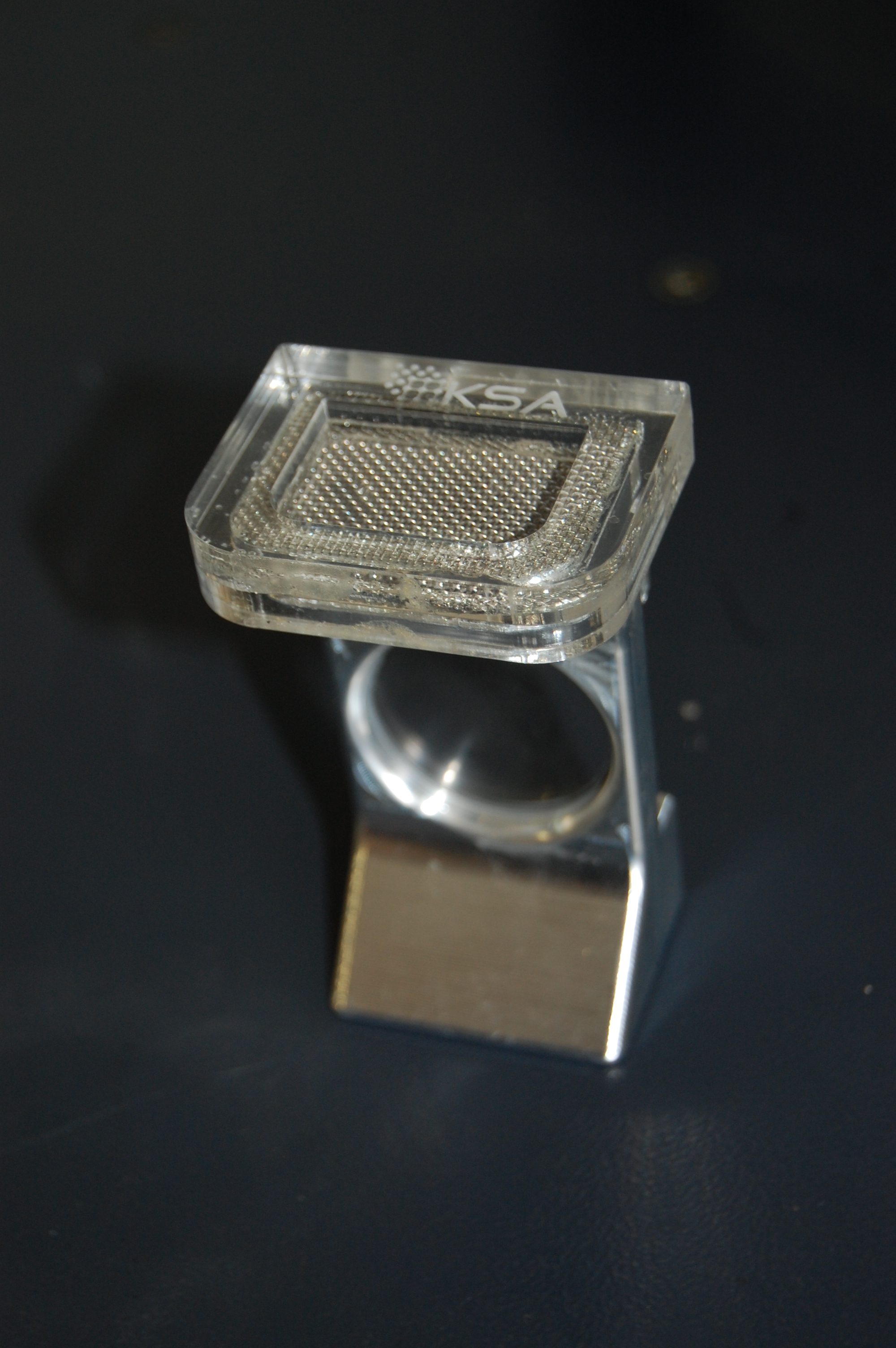
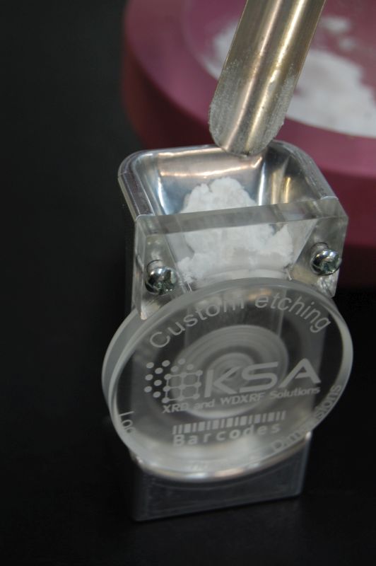
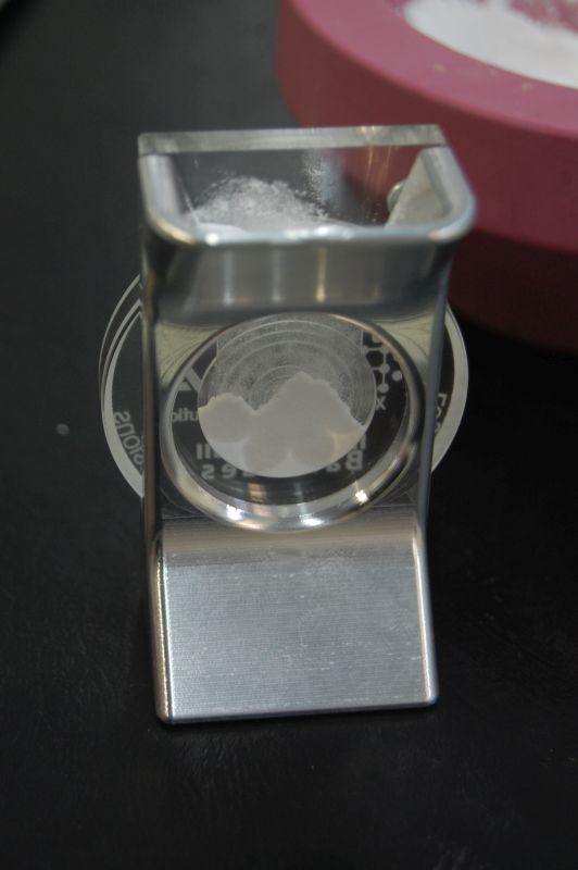
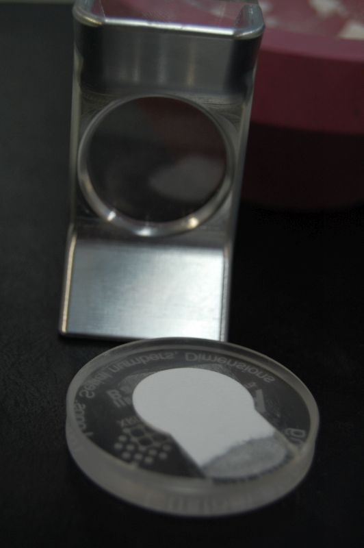
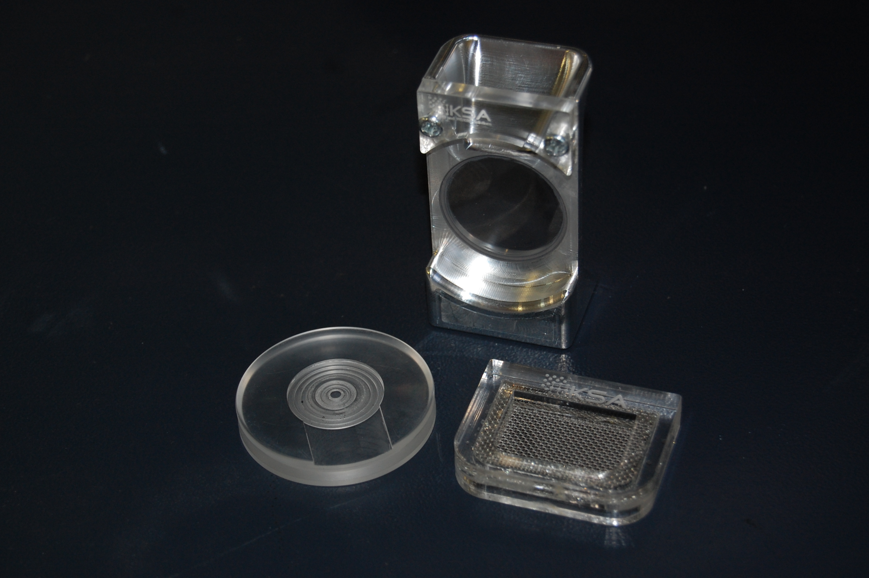
XRD sample prep is like a box of chocolates. You never know what you’re going to get… So many materials are fluffy or sticky that even after fine grinding, it’s common to have some clumps that just don’t want to break up. This became a problem for one of our clients using our side-loading tool so they added a piece of mesh to the mouth of the funnel. Their next order included a request for some type of removable solution for this so we mounted some coarse mesh in an acrylic frame that sits nicely on top of the funnel and makes it very easy to sift through sample material as it’s being loaded. We love these so they’ll be an option on all future orders!


Clay minerology is a fascinating subset of the common powder XRD applications we work with. It seems like every lab has their own unique way of handling the various challenges presented by clays and hence the need for myriad solutions. Most recently we worked with a lab which processes large volumes of clay samples in their various Bruker XRD system and they needed a sample holder that would accommodate their 26 x 26mm glass slides on which their clays are mounted. The solution was a custom holder with a square recess for the plate which is simple enough, but in cases like this, one must take into account the real-world challenges of machining as well as usage. The edges and corners of the glass slides are the least precise part of the plate so the holder has a “moat” around the perimeter to eliminate binding or shifting due to those issues. The center is also relieved as this area does almost nothing to improve precision of the mount, but will cause dramatic displacement errors if any debris gets between the holder and plate in that area.

These were a little time-consuming to make, but the end result worked beautifully. The client was very happy and we’re happy to have another design to offer to other labs with similar needs.


You can never have enough sample holders no matter what machine you’re running. We’ve certainly found this to be true at Texray so we always try to keep a large number of them on-hand. KS Analytical Systems has always made one-off and custom sample holders for the Bruker instruments, but we’re now offering the standard PMMA powder holders as well at significant cost savings over the OEM version. The standard holder (25mm x 1mm deep well) is priced at $55 with bulk discounts starting at 20 holders.

Custom well depths, diameters, grooved floors, side-loading and zero-background versions are available.

Our PMMA holders are compatible with Bruker D8 Focus – D8 Advance (single, FlipStick autosampler, 90-position autosampler), D4 Endeavor and D2 Phaser (single only) systems. D500 and D5000 instruments can also use these holders.

We’ve brought the complete manufacturing process in-house to give us the freedom to make the custom designs our customer have always asked for. This includes custom laser etching. Company logos are a common request, but we’ve also started serializing sample holders on request. At Texray, we even etch them with barcodes for tracking samples through the data collection process.

The pictures below show a custom funnel tool for filling side-loading sample holders. The tool is machined from billet aluminum with an acrylic window on the funnel to make it easier to gauge fill level. The funnel itself is polished and the viewing plate which allows the users to see when the sample well is full is made of sapphire crystal for maximum scratch resistance.

One of the fundamental facts of lab-based X-ray production is that our x-ray tubes emit much more than the pure KA1 lines we rely on for material characterization and quantification. Most XRD users are familiar with techniques and hardware for the reduction or elimination of KB1, W LA1 and Bremsstrahlung, but take for granted the inseparable pair of KA1 and KA2 (referred to as the “doublet”). Luckily for us, these energies are present in strict proportion such that we can factor their paired presence into most XRD analysis to the point that one might barely notice their effect. However, the fact remains that we will see peak broadening at lower angles and completely independent additional peaks at higher angles due to this superfluous discrete emission.

Separating the doublet cannot be accomplished electronically or through absorption/attenuation such as might be effective for KB1 energies. It must be done in the primary-beam with an additional diffraction event. Primary-beam monochromators are generally classified by the number of diffraction events required for a photon to pass completely through the device. Single-bounce, 2-bounce and 4-bounce geometries are common with the latter providing the best energy resolution allbeit the lowest intensity (photon flux). My limited experience suggests that while the single-bounce models retain enough intensity to have some application in powder XRD, the others are relegated to HR-XRD applications such as XRR.

The alignment for any of this hardware is not for the faint of heart as it begins with coarse adjustments using fluorescent screens in the beam path. This was essential for us given how dramatically misaligned the monochromator had become after so many attempts to bring it back into operation. We actually needed our SDD system to verify that we were tuning for Cu KA1 energy rather than the KB1 emissions because some of the most basic aspects of the alignment had pushed way beyond their intended position.

Along the way we built ourselves a motorized remote adjustment tool which we’ll return to the user as small adjustments are required on a regular basis with this kind of monochromator to retain maximum intensity. It’s quite useful and even versatile enough to allow for the adjustment of multiple control knobs.

One final note regarding intensity. It’s easy to get excited about energy resolution like this, but bear in mind that we’re looking at ~20x reduction in intensity due to the inherent losses involved in the primary diffraction event. This data was collected at 10x the normal speed and at half the normal 2Theta step increment so it looks very good, but one would need a compelling reason to slow their data collection this much.

Another side effect of performing your energy discrimination in the primary beampath is that other issues such as fluorescence effects (incident x-rays exciting elements in the sample causing high background intensities) are harder to avoid than they would be with a diffracted-beam monochromator. The 4x reduction in intensity inherent in the diffracted-beam monochromatization makes it a poor choice to eliminate these effects when the incident intensities are already so low. We recommend energy-dispersive detectors such as our SDD-150 to eliminate extraneous energies without sacrificing net intensity. We’ve also worked with the Bruker LynxEye XE-T detector which has a very high energy resolution compared to other position sensitive detectors (PSD). Contact KS Analytical Systems for more information on these options.

Over the past 15 years, Barnett Shale has become a major resource for natural gas in Texas.  Being located in the North Texas region, it is easy to see the boom of drilling rigs and wells popping up in the suburban and rural areas between Ft. Worth and Denton.  Collaborations between Geologists at universities and major oil companies have put a large amount of research into characterizing shale.  In 2001, Środoń et al. published a journal article in Clays and Clay Minerals that discussed the importance of sample preparation for sediments, such as shale, to be analyzed using X-ray diffraction.

Powder X-ray diffraction is the preferred and best technique to identify and quantify mineral compositions in geological materials such as rocks, sediments, and soils.  Sample preparation and loading are two important factors for accurate quantitative XRD analysis using Rietveld refinement.  Proper sample grinding and using a side-loader or backside loader are common practices to avoid preferred orientation.  At Texray, we have a variety of sample holders for different applications, and we can even custom build holders for those random parts.  However, in this study we wanted to see for ourselves the effect of sample grinding and particle size, and also we wanted to test out our new McCrone Micronizing Mill.  We already knew what the results would be from experience and previous work by Środoń et al., 2001 and Klug and Alexander, 1974, but this was a fun experiment to try with shale.

Shale rock from the North Texas region

Shale Rock from the North Texas region

The rocks (pictured above) were broken up into smaller pieces using a mortar and pestle, and then half was transferred to the McCrone mill for wet grinding and the other half we continued to grind manually using the mortar and pestle.  By the way if you are running out of bench space in the laboratory and are looking for a mill, I highly recommend the McCrone Micronizing Mill because it takes up very the little space and it’s capable of grinding below 10 μm in less than 10 minutes.  After grinding, we loaded the powder samples into a backside loader and analyzed them using a Bruker D5000 X-ray Diffractometer.

Shale XRD Pattern

XRD pattern of Mortar & Pestle Ground Shale (blue) vs McCrone Mill Ground Shale (red)

In the XRD pattern shown above the main differences you will notice between the two grinding methods are peak intensities and a small 2-theta peak shift.  Both of these differences are effects related to particle size distribution and sample loading.  Wet grinding the shale in a McCrone Mill creates smaller uniform particles (~5μm), therefore when loading the sample into holders the powders pack easier and tighter creating a denser layer of material for the X-rays to penetrate, hence higher peak intensities compared to manual grinding.  Sample preparation is one of the most important aspects to quantitative XRD because of preferred orientation and sample displacement.  In order to reduce user error such as, induced preferred orientation, it is essential we learn from previous research and take the proper steps to prepare samples.  The ICDD is a great source for free literature on applications involving XRD and XRF.  We will be posting more discussions on sample preparation and applications in the future.