The Ultimate Power Protection Solution: UPS Designs for Bruker AXS Systems by NXT Power

KS Analytical Systems is proud to present a new, total UPS solution for clinical and diagnostic laboratory systems, specifically designed for Bruker AXS, by NXT Power. The Integrity Max & Pro UPS is a complete power protection solution providing isolation, power conditioning, and backup protection for Bruker AXS systems. Regardless of input fluctuations, the Integrity […]
Rigaku D-Max zero background sample holders
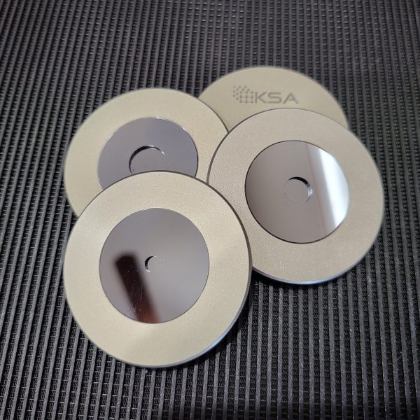
This project started with a phone call several months ago from a Canadian government lab. They had a very specific application so the design was driven much more by their requirements than most zero-background holder designs. A 1mm deep well with a zero-background plate at the bottom was a key specification. We tried several options […]
Bruker D4 Endeavor on its way out

The D4 Endeavor fits in a unique niche in the XRD world. It’s basically the same thing as a Bruker D8, but built into a very compact cabinet with a large autosampler on top. These machines see heavy use in the cement, pharmaceuticals, and aluminum industries among others. Today we have one headed out to […]
Fully digital autosampler install
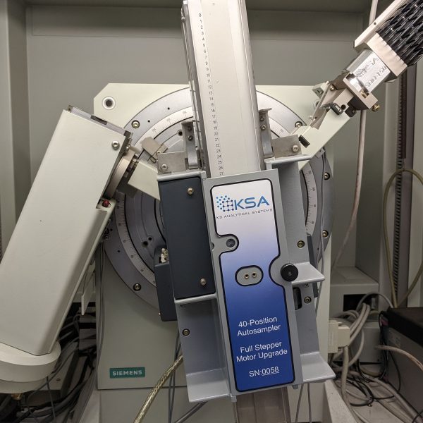
Another fully-rebuilt, digital autosampler out in the wild. This one is on a system that already has one of our Si-Drift Detectors and an awesome ICDD Jade Pro/PDF-4+ software package. We’ve got all the fancy new hardware at our in-house lab, but when we need the absolute best data, this is our goto configuration.
Sample holders for Rigaku Miniflex systems
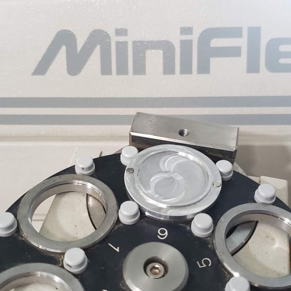
KS Analytical Systems now offers custom sample holders for Rigaku Miniflex systems. The 6-position autosamplers and rotation stages (and even some fixed stages) make use of the magnetic disk design for holding powder without taking up much extra room in the diminutive benchtop. Top-loading Rear-loading Zero-background